Patella laxation is also called loose knees. Most the time if your dog or puppy has them the vet will grade it, The vet grades them from a 1-5, 1 being a little loose and 5 being the worst. Toy breeds are some of the most common breeds for this issue. That is why is is so important that breeders have their adults tested and Registered with the OFA showing their adults don't have this problem. This is a issue that can be congenital, or could also be how the puppy was raised. If while the puppy is growing and maturing if they don't have a solid ground to walk on this could cause Patella laxation. If this is the case some times the knees can tighten when they get on solid ground. But if it is congenital they will most likely just get worse as they grow and mature.
Below you will see some great information about and explaining patellar luxation.(USED with permission)
Medial and lateral patellar luxation Dislocating kneecap, luxating patella, loose knee, trick knee
AffectedAnimals: Dogs, cats, humans
Patellar luxation can be a congenital condition or an environmental condition in which the kneecap, or patella, dislocates outside of its normal trochlear groove. Dislocation, clinically referred to as luxation, can occur on either the medial, or inside surface, or the lateral, or outside surface, of the knee. There are varying degrees of patellar luxation that are graded depending on whether the patella is intermittently or constantly luxated. This abnormal displacement of the kneecap results in pain, cartilage damage, and arthritis. There are varying degrees of severity of this disease, and surgery may be needed.
 Courtesy of: Gale Mueller
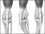
The knee on the left shows the normal position of the patella. The center knee shows luxation of the patella to the medial side. The knee on the right represents luxation of the patella to the lateral side.
Courtesy of: Gale Mueller
The knee on the left shows the normal position of the patella. The center knee shows luxation of the patella to the medial side. The knee on the right represents luxation of the patella to the lateral side.
It is much more common for the kneecap to ride on the inside than on the outside surface of the knee. This condition typically affects small and miniature breeds such as the poodle, Pomeranian, Chihuahua, Schipperke, Bichon Frise, and pug. It is also seen in the cocker spaniel, golden retriever, Labrador retriever, and mixed breeds.

Outside dislocation of the kneecap, or lateral patellar luxation, occurs more often in large breeds such as the Great Dane, Saint Bernard, and Bernese mountain dog. Lateral luxation is often accompanied by malformation of the femur, the large bone in the thigh. In these cases the prognosis is more uncertain, as major limb reconstructive surgery may be needed.
Generally, the prognosis is better when corrective surgery is performed early in the course of the disease; many of the malformations in the bones and joint occur over time and can be prevented with early correction.
Lameness that is often intermittent, and may be unilateral or bilateral; thick, swollen stifles; pain on range-of-motion; crepitus; palpable luxation; inability to jump or walk normally; medial displacement of quadriceps muscle group; lateral bowing of the distal third of the femur.
Intermittent or consistent lameness; bowlegged stance; reluctance to walk or jump; occasionally holding a rear leg out to the side when walking.
Medial patellar luxation, or MPL, is a very common disease of small and miniature breeds in which the kneecap occasionally rides on the inside of its normal groove. Primarily congenital, although occasionally acquired through trauma, MPL causes lameness in one or both rear limbs. The degree of lameness is determined by the severity and duration of the disease, as well as the extent of existing arthritis.
Patellar luxation is graded on a scale from I to IV, with IV being the most severe. The disease can progress from the less severe to more severe grades over time. The more severe forms are often accompanied by malformation of the femur and tibia, as well as varying amounts of arthritis.
Dogs are frequently presented to a veterinarian for intermittent lameness, often because it is becoming more frequent or severe. When the patella, or kneecap, pops out of its normal trochlear groove, the dog feels pain, and owners may report a hitch in the gait. The dog will frequently extend the knee out from the body in order to get the patella to pop back in to the trochlear groove. As MPL progresses, the structures that hold the patella in place become looser, and thus the problem becomes more frequent. This dislocation causes pain, and as the frequency increases, so does the lameness.
Unfortunately, many of the severe Grade III or IV cases go unnoticed for months or years because the affected animals are usually miniature breeds that are often carried much of the time by their owners. Their inability to jump or straighten out their hind legs may go unnoticed because of their small size and sedentary lifestyle.
Lateral patellar luxation, or LPL, is less common than MPL and occurs when the kneecap occasionally rides on the outside of its normal groove. It, too, can be congenital or acquired, with the congenital form again being more common. While it can occur in any dog, it is more common in large and giant breeds. LPL is frequently accompanied by malformation of the femur and/or tibia. The disease can produce marked lameness and progress to crippling arthritis. Because of the accompanying bony malformations, extensive surgery may be required to correct this problem.
The examining veterinarian will often make a diagnosis from a physical examination and history. However, x-rays are needed to determine the degree of arthritis, and evaluate for any malformation of the femur and tibia, the two major bones in the leg, which are joined together at the knee.
The prognosis for a Grade I patellar luxation is very good. These dogs may not need surgery. However close observation for signs of worsening is important. If surgery is indicated and performed early on, most animals regain normal functionality.
The prognosis for Grades II and III depends on how much arthritis and malformation have occurred. If caught and treated early, both have a good to excellent prognosis. If there is significant bony malformation or arthritis, the prognosis is guarded to fair.
The prognosis for Grade IV patellar luxation is guarded. Most of these animals have moderate to severe bony malformations and significant arthritis. If correction is performed, it is important to initiate early physical therapy to help restore function.
The congenital condition is probably genetic in nature, and as such, affected animals should not be bred. Trauma or injury can also cause patella luxation.
Treatment involves replacing the kneecap into the groove, and preventing it from popping in and out. The following procedures can be used alone or in combination as necessary to maintain the proper function of the knee.
Tightening the joint capsule, known as imbrication, is done on the opposite side of the luxation to prevent the kneecap from having enough slack to pop out of the trochlear groove. Thus a medial patellar luxation is treated with a lateral imbrication, and vice-versa. Additionally, the joint capsule can be loosened on the side of the luxation; this is called a release incision. This procedure relieves the tension that the joint capsule is placing on the patella, thus allowing it to ride in the trochlea.
In severe cases a synthetic suture is sometimes necessary to keep the kneecap in place. This suture is placed on the side opposite the luxation, and goes from behind the femur to the patellar tendon. It also prevents the kneecap from popping over to the other side.
Deepening of the trochlear groove, or trochleoplasty, can be accomplished with a variety of techniques. A chondroplasty technique involves cutting out a taco-shaped wedge of cartilage, removing a small portion of bone beneath it, and then replacing the cartilage. The result is a deeper groove. This procedure can only be performed on very young dogs, because their cartilage is thicker.
Trochlear recession involves cutting out the cartilage and bone in such a way as to create a deeper trough. This trough will then fill in with scar tissue over time. Because this scar tissue is not as good as cartilage for joint function, this technique has given way to others that attempt to preserve normal cartilage. It can, however, be useful in carefully selected cases.
Wedge recession creates a taco-shaped piece of cartilage and underlying bone. Then, the bone below the wedge is removed and the wedge is replaced, forming a deeper groove. Block recession is identical in principle to wedge recession, except that a rectangular piece of cartilage and bone, rather than a wedge, is removed.
The kneecap attaches to the lower leg via its patellar tendon at a bony site called the tibial tuberosity. Many times this site forms abnormally on the inside, as with MPL, or on the outside, as with LPL. In this procedure, the surgeon moves the tibial tuberosity back into proper alignment and secures it in place with a pin or wire. Realigning the joint, kneecap, and tendon prevents dislocation from reoccurring.
In severe cases, with malformation of the tibia or femur, corrective bone cuts known as osteotomies may be required.
Early detection and correction is the best way to prevent severe lameness and dysfunction. Breeding affected animals should be discouraged; however, the disease is so prevalent in some breeds that this may not be practical.